
At the Helios Klinikum Berlin-Buch, catheter ablation was performed using a cardiac MRI. For the first time, a team of cardiologists has paired the standard procedure of catheter ablation with a cardiac MRI. In the future, this new method promises to improve the treatment of complex cardiac arrhythmias through real-time imaging and more precise treatment of heart tissue. The head of the Heart Rhythm Center Berlin/Brandenburg at the Helios Klinikum Berlin-Buch Dr. med. Michael Wiedemann explains the innovative process in an interview.
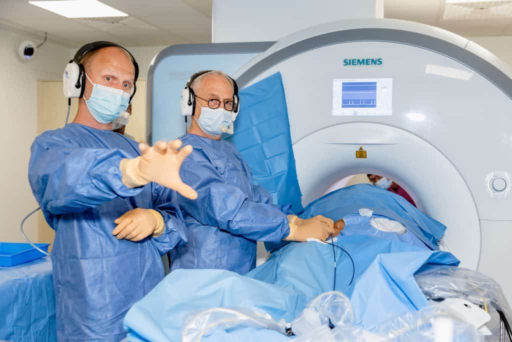
Photo credit: Kareen Kittelmann | Helios Clinics
What are the prerequisites for this particular procedure?
Dr. Wiedemann: “Due to our excellent experience in the team and interdisciplinary cooperation as well as our special equipment of a cardiac MRI and additional instruments, we were able to perform this MRI-guided catheter ablation for the first time in Berlin. For the surgical procedure with MRI imaging, cardiac catheters are required, which are not attracted to the magnetic MRI machine. That’s why we used plastic catheters with a gold tip and two electrodes, which were specially developed for this application. This makes us one of the first ten cardiology centres in Europe to be able to implement this method.”
What is a cardiac MRI?
Dr. Wiedemann: “Our state-of-the-art cardiac MRI is located on the garden floor of our hospital. This “cardiac MRI” makes moving images of the heart anatomy in real time. The procedure is radiation-free and enables doctors to obtain very precise examination results, as the MRI images represent the individual structure and function of the heart very accurately. For example, special features such as complicated bulges of the fabric are displayed. In this specific case of MRI-based catheter ablation, we precisely treated atrial flutter, a special form of cardiac arrhythmia, in our patient and were able to look at the MRI images during the procedure. My colleagues from our cardiac MRI department benefit greatly from the leadership of Prof. Dr. med. Jeanette Schulz-Menger, who has already received several awards for her excellent research work.”

Photo credit: Thomas Oberländer | Helios Clinics
What is atrial flutter?
Dr. Wiedemann: “In typical atrial flutter, the electrical signal in the heart is disturbed. The signals come off the path and “circle” in the right atrium. This so-called excitation circle is the same for all patients. As a result, the atria can no longer pump properly: they “flutter” and are excited up to 300 times per minute. In Germany, the diagnosis of atrial flutter and especially atrial fibrillation is the second most common reason for hospitalization. Atrial flutter is usually treated by catheter ablation.”
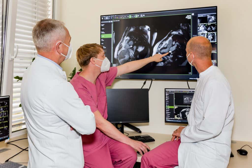
Photo credit: Kareen Kittelmann | Helios Clinics
What is catheter ablation?
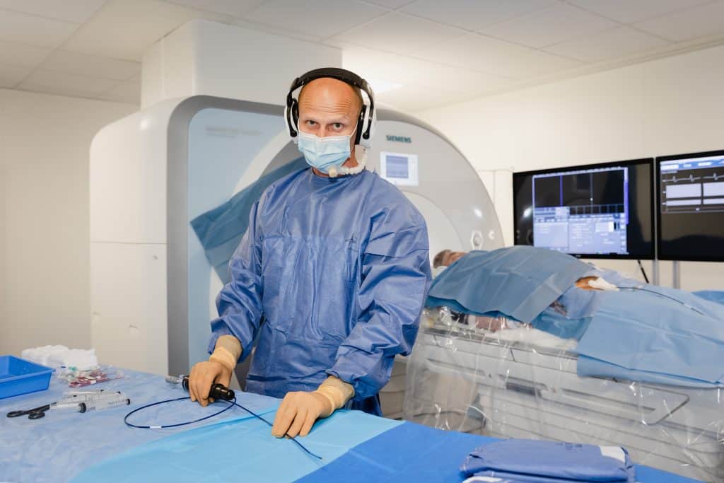
Dr. Wiedemann: “With a few exceptions, catheter ablation is the standard procedure for the treatment of cardiac arrhythmias. As an imaging method, fluoroscopy with X-rays is usually used here and combined with a navigation method. In the minimally invasive procedure, catheters, flexible plastic tubes, are pushed over the inguinal vein to the heart and placed in the heart. The ablation catheter selectively obliterates heart muscle tissue with heat generated by high-frequency current. Another method is sclerotherapy by cold. The sclerobed cells can no longer transmit the electrical signal and – as in our case of atrial flutter – the circling excitations are interrupted. The signal in the heart can be transmitted again in an orderly manner and the heart rhythm is intact again.
How many cardiac MRIs, catheter ablations and electrophysical examinations are performed on average per year at Helios Klinikum Berlin-Buch?
Dr. Wiedemann: “In our four highly specialized cardiology laboratories, we perform special catheter-based cardiac examinations for diagnosis and therapy. Two of these laboratories are specially equipped for the treatment of cardiac arrhythmias. Around 1000 treatments such as ablations and electrophysical examinations take place at Helios Klinikum Berlin-Buch every year. In our cardiac MRI under the direction of Prof. Dr. med. Jeanette Schulz-Menger also examines around 3,000 hearts per year.”
What are the advantages of this MRI-based catheter ablation procedure?
Dr. Wiedemann: “In the future, complex arrhythmias, especially life-threatening tachycardia conditions, will be the focus of this treatment method. MRI imaging can not only visualize anatomical features of the heart in real time, but also visualize scarred changes. Frequently, these scars cause very different, sometimes life-threatening cardiac arrhythmias. With the help of MRI, it should be possible to ensure optimal therapy planning and implementation. This is an important advantage over the classic “X-ray”, a fluoroscopy-assisted catheter ablation. Although there are already other navigation systems for heart interventions that enable 3D planning and are also used in surgery, they cannot display the treatment and results in real time as with MRI. In addition, the procedure does not result in any X-ray exposure for patients or medical staff. Further research and technical advancements are needed to bring these benefits into practice. We are looking forward to this interesting work.”
What is rhythmology?
Dr. Wiedemann: “Rhythmology is a special field of cardiology and deals with the diagnosis and treatment of cardiac arrhythmias. Rhythmology is the science of electrical excitation in the heart. In order to detect and treat disorders, we use the ECG or a cardiac catheterization. Standard therapies then are, for example, catheter ablation for tachycardia or a pacemaker for a too slow heartbeat. We carry out diagnostic examinations and procedures in our state-of-the-art cardiac catheterization laboratories with all the technical requirements for special catheter-based cardiac examinations.”
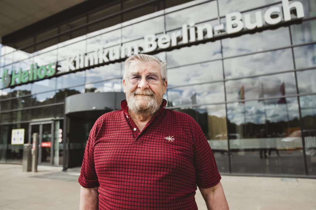
“My goal is to be 100.”
Gerd-Heiner Pippis (70) is in good spirits after innovative procedure with cardiac MRI
After a routine examination by the family doctor, Gerd-Heiner Pippis is sent to the hospital. The physicians from the Helios Klinikum Berlin-Buch diagnose a severe cardiac arrhythmia, a so-called ventricular flutter. The sinus rhythm in the heart is out of sync and the electrical signal in the heart circles in the atria and makes them flutter because the heart can no longer pump properly. At first, the pensioner from Berlin-Buch is a bit shocked. “I never had any symptoms: no tachycardia, no attacks of weakness,” he says.
Cardiac MRI-guided catheter ablation
Gerd-Heiner Pippis comes to the clinic at the right time. In order to bring the heart back into “tact”, he not only uses the standard procedure of catheter ablation, but also a completely new procedure: an ablation with simultaneous cardiac MRI. His attending physician, Dr. med. Michael Wiedemann, Head of the Heart Rhythm Center at Helios Klinikum-Berlin-Buch, explains the procedure. “MRI-guided catheter ablation enables very precise examination results, as the cardiac MRI images represent the individual structure of the heart very accurately. In this way, even special features, such as a complicated tissue structure, can be displayed and taken into account during the procedure,” explains the doctor. “This procedure could be carried out for the first time in Berlin by the Bucher cardiologists due to special surgical instruments. This makes us one of the first ten cardiology centers in Europe to be able to implement this method,” adds Dr. Wiedemann.
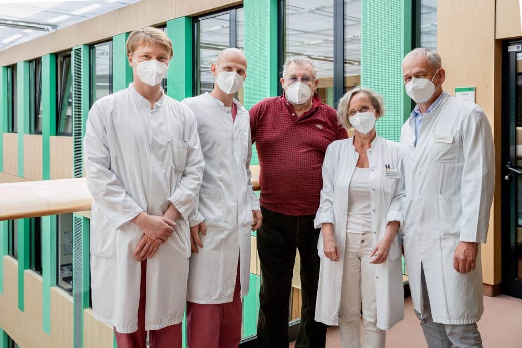
Photo credit: Kareen Kittelmann | Helios Clinics
Already the second operation on the heart
About seven years ago, an ablation of the pulmonary veins was successfully performed on the patient Gerd-Heiner Pippis, which did not discourage the native of Trier or prevent him from remaining active.
The former business computer scientist likes to travel with his “baby Harley”. After the effective treatment of his “atrial flutter” with the help of MRI-guided catheter ablation, Gerd-Heiner Pippis was able to ride home on his motorcycle the following day. “Now my sinus rhythm is fine and I can enjoy my life again.” Now he feels really good: “My goal is to turn 100,” says the sociable 70-year-old.
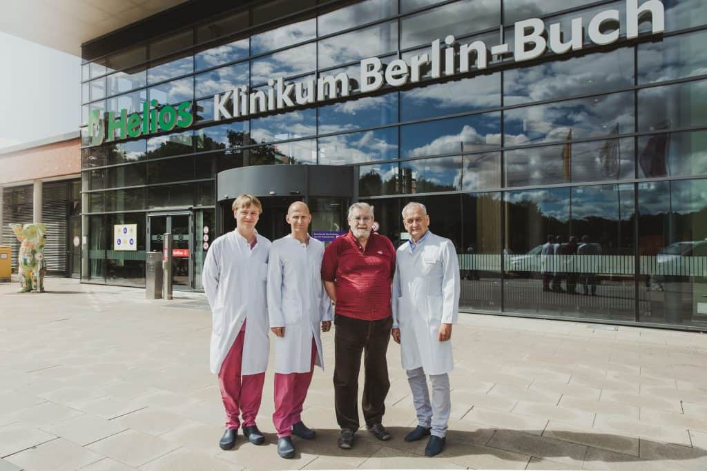
Photo credit: Kareen Kittelmann | Helios Clinics
Orginial press release from:
Helios Klinikum Berlin-Buch “First Time in Berlin: Catheter Ablation Performed with MRI Imaging” [Press Release] Retrieved from https://www.helios-gesundheit.de/kliniken/berlin-buch/unser-haus/aktuelles/detail/news/erstmals-in-berlin-katheterablation-mit-mrt-bildgebung-durchgefuehrt/