At the Cardiology Department of Amsterdam UMC, location AMC, they are doing something groundbreaking. For about a year now, patients with certain cardiac arrhythmias have been able to come here for an innovative treatment: ablation while lying in an MRI-scanner. We visualized how such an interventional Cardiac MRI (iCMR) works.
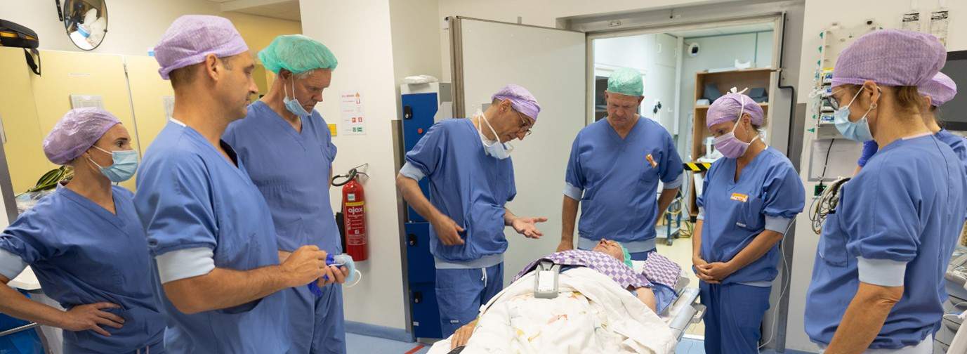
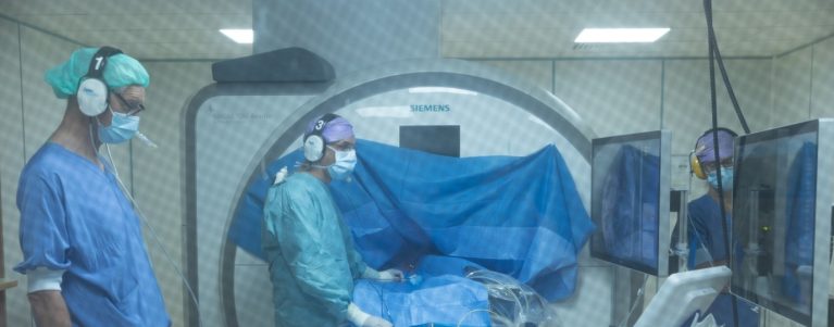
The treatment is done under anaesthesia. Before the patient is put to sleep, they are given an explanation and there is a final check. The team also introduces themselves. There are quite a few people around the patient. More than with a ‘regular’ ablation. To be able to carry out this new form of treatment, various employees work together. Several lab technicians, an anaesthesiologist who provides the anaesthesia, several nurses and two cardiologists. Marco Götte (imaging cardiologist) and Cor Allaart (cardiologist-electrophysiologist) started researching the possibilities of iCMR in patients with cardiac arrhythmias about 10 years ago.
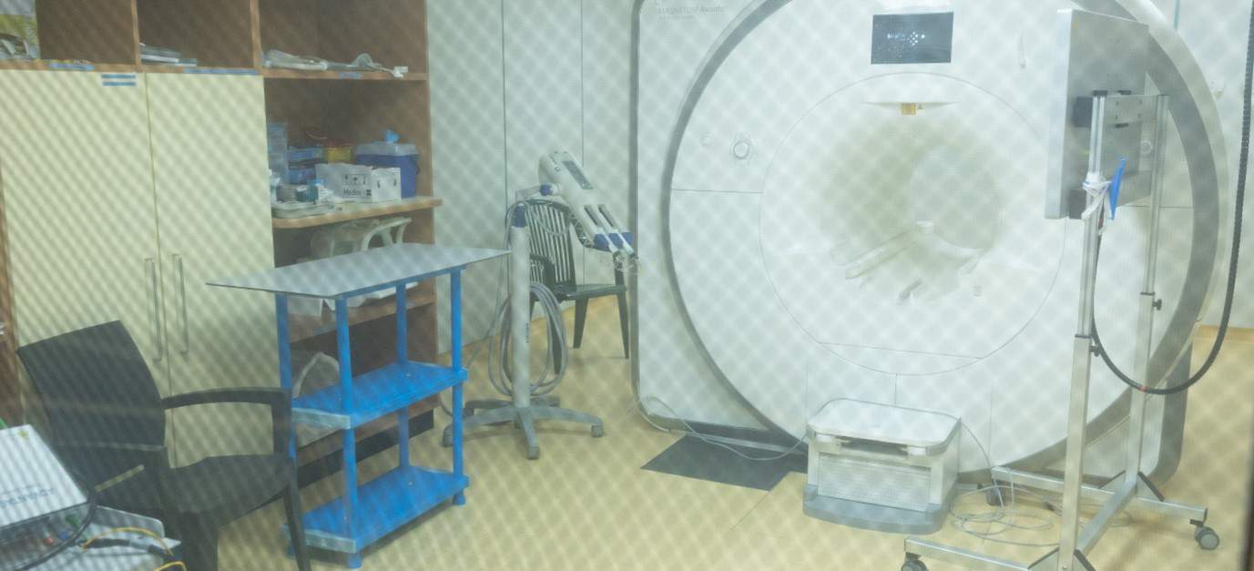
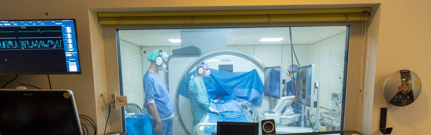
In the room where an MRI scanner is located, nothing made of steel, or any other magnetic material, may be used. The MRI scanner itself works like a large, very powerful magnet. But because this method of treatment is new, there is still very little equipment that is suitable. So, they now use, for example, a plastic table.
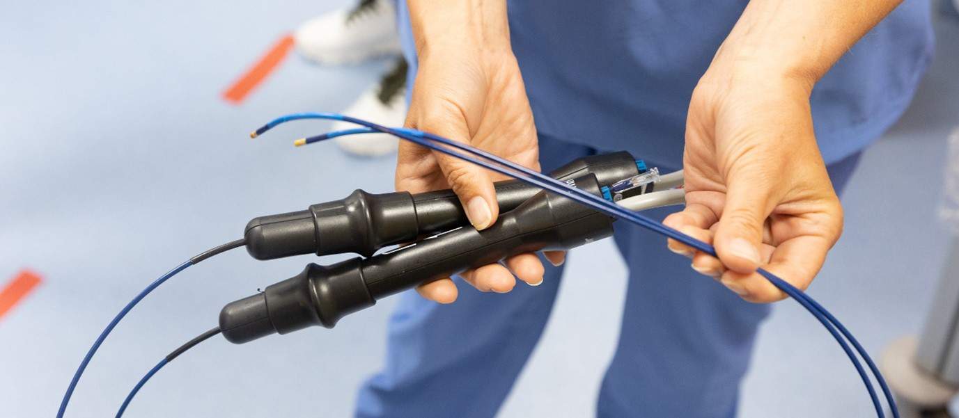
An ablation is done using two catheters. Thin, flexible tubes that go through the groin to the heart for this treatment. At the end of one catheter is a tip that gets hot. This creates the ‘scars’ on the inside of the heart. Below the tip is a kind of reflector that lights up through the MRI scanner. This makes it possible to see exactly where the catheter is on the screen. Everything is monitored with the other catheter. The cardiologist-electrophysiologist controls the catheters and can thus treat the patient externally.
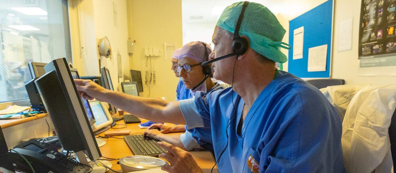
Only the cardiologist-electrophysiologist and a nurse are in the MRI scanner room with the patient. The rest of the team is in the control room. They monitor the screens and communicate with the electrophysiologist via headsets.
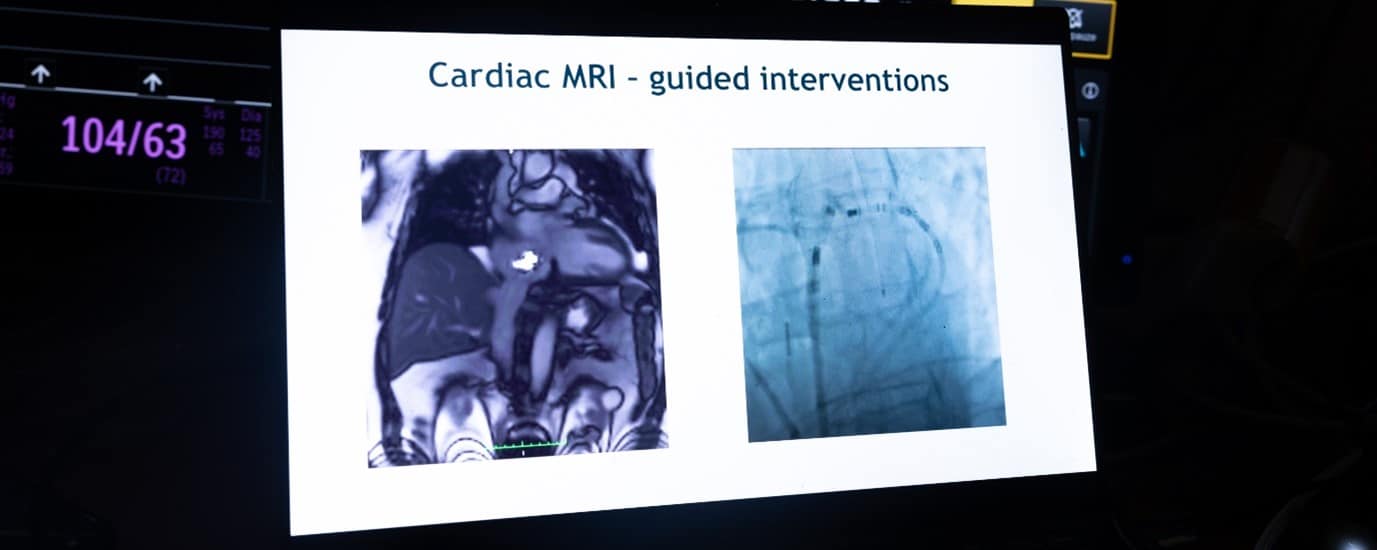
What makes this method of ablation so special and so important is that the team can see what is happening much more precisely. And that the electrophysiologist can steer much more precisely. An MRI can provide images from all sides/directions: for example, a cross-section of the body from the front, from the side and from above. Everything in and around the heart is clearly visible. An X-ray system, with which these treatments are normally done, only gives a 2D image. This treatment is also much nicer for the patient. So, the chance that the patient needs to be treated again is much smaller.
For now, only the ‘simple’ cardiac arrhythmias can be treated with the aid of an MRI scanner. This will remain the case in the near future, as the technology isn’t yet ready to perform more complicated procedures. But if all goes well, that will be different in 2 years. Then the new Heart Center at the AMC location will be ready, and a newly developed MRI scanner will also be installed. Especially for these treatments. New special software has also been developed. This will hopefully make it possible in the future to also perform more complicated treatments on the heart. As far as this team is concerned, the road to that goal is clear.